
Journal of Cardiovascular Magnetic Resonance,
Imaging study
Help us to complete the world's largest imaging study
The UK Biobank imaging project aims to collect brain, heart, and abdomen scans from 100,000 participants. Thanks to the help of our participants, we have collected imaging scans from over 60,000 participants to date. Performing imaging at this scale is unprecedented and will allow researchers to identify associations between lifestyle, genetic factors, imaging-derived phenotypes and how these affect disease risk.
We take very seriously the health and safety of our participants and staff alike, and put in place strict measures to ensure that our imaging centres are safe. We routinely review our operational practices to reduce the risks to safety and to prevent the transmission of infections.
To help with this, please do not attend your imaging clinic appointment if you have an infection which might affect or be transmitted to those around you, for example a respiratory infection (such as COVID-19 or a cold), influenza or shingles.
Advancing the diagnosis, prevention and treatment of disease
The UK Biobank imaging study is enabling scientific advances in imaging and analysis, opening up new ways to diagnose, treat, and potentially prevent diseases like dementia, heart disease, and cancer. It will capture a vast amount of images of the human body during both good and ill health. Combined with the genetic and lifestyle data already held on half a million participants, it will shine a light on why one person develops a life-altering disease when others do not.
Imaging videos on our YouTube Channel
The UK Biobank imaging study is the most ambitious project to take pictures of the inside of the body ever undertaken.
We are scanning the brains, hearts, bones and abdomens of 100,000 UK Biobank participants – and we are already half way there.
If you have taken part – then thank you so much. If you have not yet been invited, do not worry as we plan over time to invite all UK Biobank participants to be scanned.
Who is funding the study?
The £43 million study has been funded by the Medical Research Council, Wellcome Trust, and the British Heart Foundation (BHF), with a further £10 million from Dementia's Platform UK to re-image 10,000 participants.
Related Publications
Wellcome Open Research,
Impact of detecting potentially serious incidental findings during multi-modal imaging
Lorna M Gibson
How will I be invited?
We are inviting participants by email (where we have an email address) and by post. If you are interested, please call the Participant Resource Centre on 0800 0 276 276. We will take you through a short telephone questionnaire with one of our team at the UK Biobank Participant Resource Centre. This is to make sure that it is safe for us to scan you (some implants, and existing health conditions mean we cannot scan you).
Where are the imaging centres?
We have imaging centres in Newcastle upon Tyne, Stockport, Reading and Bristol. For some participants this means you will have to travel further than when you attended your original assessment. We are sorry about this (you can claim expenses). Because of the huge cost in building an imaging centre, we have based them at the centre of the largest number of UK Biobank participants in a given area.
A unique picture of health
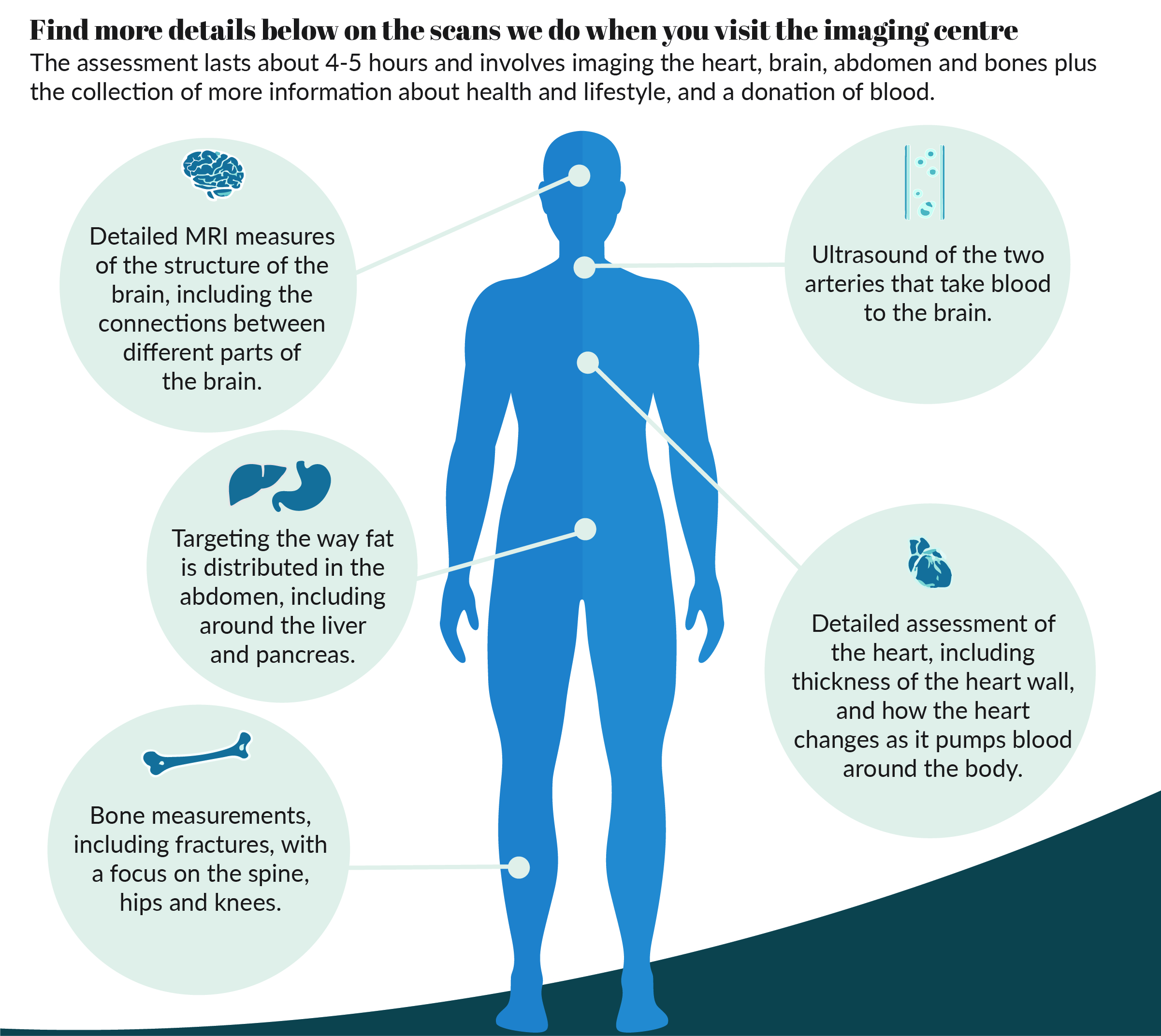
The assessment lasts around 4-5 hours and involves imaging the heart, brain, abdomen and bones plus the collection of more information about your health and lifestyle, and a donation of blood.
- MRI assessment of heart chamber diameter, the volume of blood flow, and how the heart changes as it pumps blood around the body, thickness of the heart wall and the size, shape and stiffness of the thoracic aorta, the vessel that delivers blood from the heart
- MRI measures of brain structure and function, volumes of grey matter and the mapping of major brain connections
- Dual-energy X-ray absorptiometry measures of bone density, osteoarthritic change at spine, hip and knee, fractures in the spine, and fat distribution throughout the body
- MRI measures of abdominal fat volume including in the liver and pancreas
- Ultrasound assessment of two major arteries, the carotid arteries, that run either side of the neck to the brain
What fellow participants say:
How will the images be used?
The multi-organ scans will be analysed alongside the vast data already collected from UK Biobank participants. This extra layer of data, for all health scientists to access, will give new perspectives on the best way to prevent and treat multi-faceted conditions like arthritis, coronary heart disease, Alzheimer’s disease and osteoporosis. It has already sparked new ways in which to analyse and interpret scans. Such a vast amount of data needs innovative techniques for analysis. Heart scan measurements are usually manually taken, but with over 9,000 images of the heart taken per participant, researchers have had to develop new computer based techniques, with potential benefits for research as well as for the investigation of patients in the future.
Will you tell me if you spot something wrong with my health?
The scans we do are not intended to diagnose disease. They are not designed to find any particular abnormalities and will not be routinely analysed by doctors or other specialists.
The technicians (radiographers) who do the scans will be looking at the images to make sure of their quality, rather than looking for evidence of any health problems.
Learning about fat distribution
The MRI scan will provide key information on the amount and distribution of fat and muscle mass. Combining these data with information about lifestyle, genetics and markers in the blood (like hormones, sugar and cholesterol) will hugely strengthen UK Biobank for studies into obesity and diabetes, high blood pressure, high cholesterol, cardiovascular disease and some cancers and other medical complications.
With increasing evidence to suggest that fat distribution, rather than amount of fat, is important for determining an individual’s risk of future disease, abdominal MRI provides an exciting opportunity to examine the predictive importance of particular fat depots (visceral, liver and pancreas) and the relative distribution of fat for the development of disease.
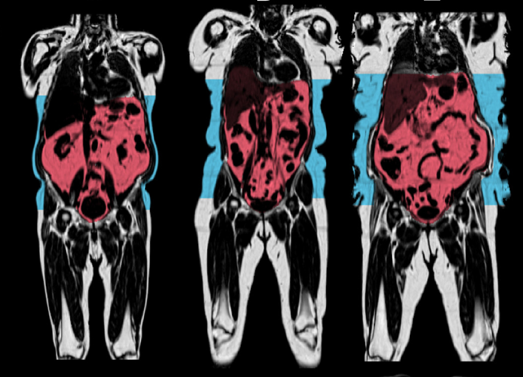
Image courtesy of AMRA -Advanced MR Analytics AB
Healthy brains
MRI scans of the brains of almost 10,000 UK Biobank participants have shown that factors that influence the health of our blood vessels, such as smoking, high blood pressure, obesity and diabetes, are linked to less healthy brains. The strongest links were in areas of the brain known to be responsible for our more complex thinking skills, and which deteriorate during the development of Alzheimer’s disease and dementia.
With the exception of high cholesterol, all of the other vascular risk factors were linked to greater brain shrinkage, less grey matter (tissue found mainly on the surface of the brain) and less healthy white matter (tissue in deeper parts of the brain). The findings show the potential of making lifestyle changes to improve brain and cognitive ageing.
Heart Monitor: can you help doctors prevent stroke and dementia?
If you are aged 65 and over you may be asked if you would be prepared to help us learn more about common heart rhythm disturbances, such as atrial fibrillation (AFib). Professor Barbara Casadei from Oxford University, who leads the heart monitoring study, explains why this is so important.
Last updated