An artificial-intelligence software analyses heart scans in seconds and allows clinicians more time to spend with their patients.
Summary
An artificial-intelligence program, developed with heart images from UK Biobank participants, gives doctors more time with their patients. The program analyses heart scans in seconds – a fraction of how long it takes clinicians to do this by hand. The software now assesses hundreds of thousands of heart scans done every year at almost 2000 hospitals worldwide.
An artificial-intelligence (AI) computer program, developed with heart images from UK Biobank participants, cuts the time it takes to analyse cardiac scans from half an hour to mere seconds. The software is now used in almost 2000 hospitals in more than 90 countries.
“It allows clinicians to spend more time with the patient rather than in front of a computer trying to analyse images,” says Qiao Wei, chief technology officer at Circle Cardiovascular Imaging, the Canadian company behind the software.
Time-consuming
It allows clinicians to spend more time with the patient rather than in front of a computer trying to analyse images.
Qiao Wei, Circle Cardiovascular Imaging, Canada
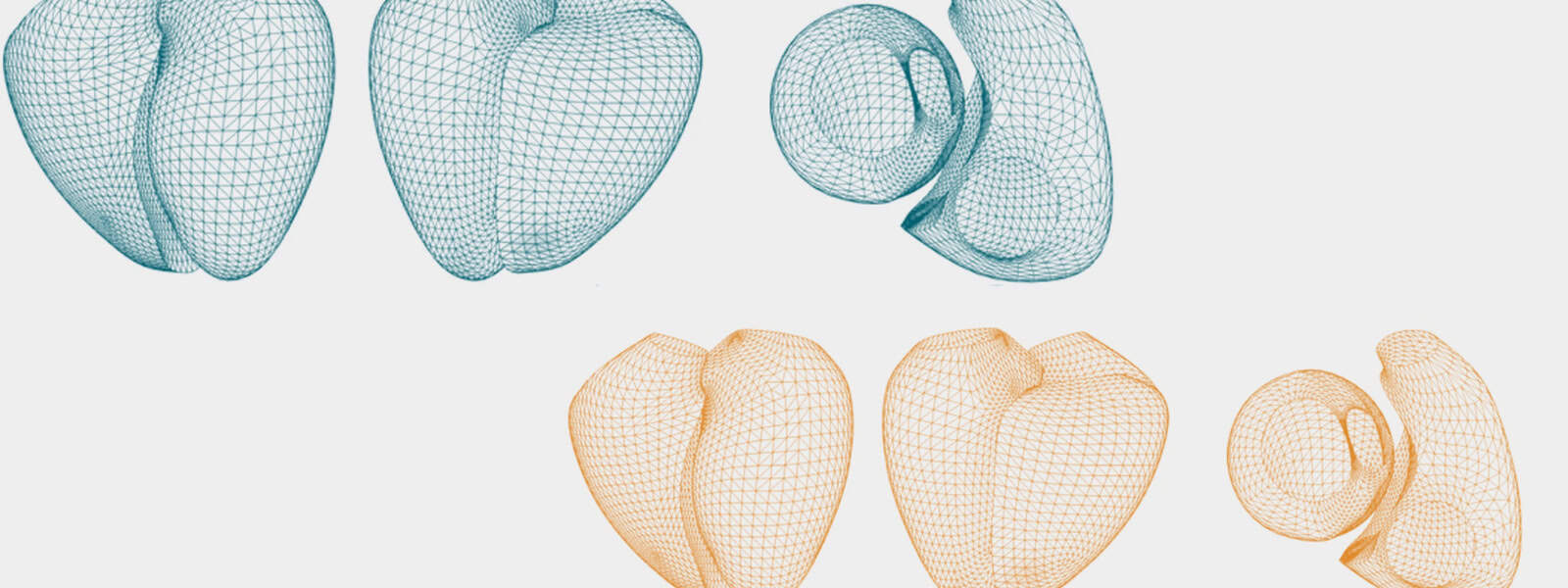
Cardiac magnetic resonance imaging (MRI) takes detailed pictures of the heart. This allows doctors, for example, to assess damage after a heart attack or to spot signs of conditions such as heart failure. More than 100,000 heart MRIs are done in NHS hospitals every year.
Before Circle’s software became available, clinicians had to manually outline the heart’s shape in MRIs. This process, called contouring, is the basis for evaluating heart health from measurements such as chamber volume.
Manual contouring is extremely time-consuming, says Steffen Petersen, a researcher Queen Mary University of London, UK, and consultant cardiologist at Barts Health NHS Trust. And it’s a source of inconsistencies, he adds: “If I’m analysing the same image twice, you probably get similar numbers, but they’re not quite the same. And it’s even worse when you have different people analysing the same image.”
An earlier version of Circle’s software cut analysis time from around an hour to 20 minutes, Wei recalls. “That’s where UK Biobank came in.”
How to teach AI about human hearts
Before UK Biobank data became available, Circle only had access to heart MRIs from a few people – not enough to train an AI program with. “If you develop an algorithm off of 50 or 100 [people], you’re basically saying, ‘This small group of people, they represent the broader public’,” Wei explains. “Software that’s trained on such a small dataset is going to fail.”

The software is now used in almost 2000 hospitals in more than 90 countries.
Circle’s AI software was initially developed with 5000 pre-analysed heart images from UK Biobank participants. The scans had been contoured manually, over the span of two years, by two experts from Petersen’s group and a collaborating team.
“Circle further developed their [software] to make it really robust in clinical settings,” Petersen says. The program now analyses heart MRIs at the press of a button. “It saves me a lot of sanity that I don’t have to contour everything anymore,” Petersen adds.
Beyond the heart
I don’t think it can be underestimated how important it was that UK Biobank created this resource of images.
Professor Steffen Petersen, Queen Mary University of London, UK
“I don’t think it can be underestimated how important it was that UK Biobank created this resource of images,” Petersen says. “UK Biobank really has been a game changer for us and the overall industry,” Wei echoes. His team is now looking at whether the images can teach an AI to recognise different diseases from heart scans.
Petersen would welcome a program that automatically analyses and flags any problems in the other organs – kidneys, lungs, spine – that are captured during a heart MRI. “This is something that I’m talking to Circle about… and maybe use UK Biobank data to start with,” he says.
Related publication
Related publication
- Circulation: Cardiovascular Imaging, September 2019


