
We’ve reached 100,000 whole body scans!
Learn more about how we’ve reached this milestone and the impact that this imaging data is having on science.
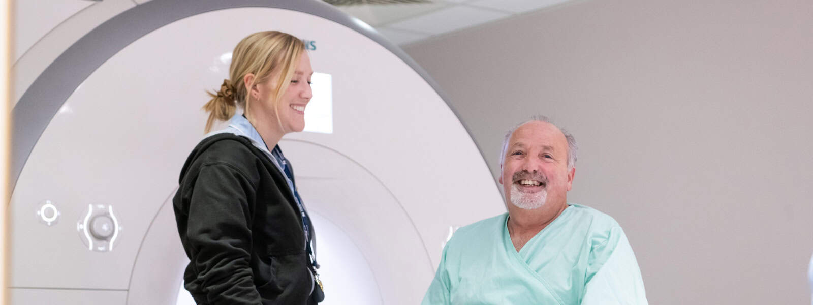
Our imaging project is the largest imaging study ever undertaken and it is transforming research into diseases like dementia, heart disease, arthritis and diabetes.
We are collecting scans of the brains, hearts, abdomens and bones of more than 100,000 of our volunteers, and asking up to 60,000 of them to return for repeat imaging 2-7 years later so that scientists can compare the first set of scans with the second.
Because of you, these imaging data are providing scientists with unprecedented insights into the health of half a million people. Our global community of approved researchers are combining these imaging data with other information, like your lifestyle habits and genetics, to understand how the diseases of ageing develop.
Take part

Be part of the world’s largest whole-body scanning project to transform the way we diagnose, prevent and treat our most chronic diseases.

Return for a repeat imaging visit to help provide researchers with information about how your body has changed over time.

Information about how to find us, parking, and travel expenses for your imaging visit.

100,000 people have been imaged as part of our initial imaging project
More than 5,000 have returned for a second time as part of our repeat imaging project
About the project
UK Biobank’s imaging project is the result of a collaboration between the government-funded Medical Research Council (MRC), Wellcome, the British Heart Foundation (BHF), and Dementias Platform UK. Additional funding to re-scan 60,000 participants is being provided by the MRC, the company Calico, and the philanthropic Chan Zuckerberg Initiative (CZI).
Will I receive any feedback about my scans?
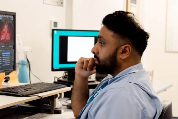
The scans we conduct are not intended to diagnose disease or find any particular abnormalities, and will not be routinely analysed by doctors or other specialists. The technicians (radiographers) who do the scans will be looking at the images to make sure of their quality, rather than looking for evidence of any health problems.

Research using imaging data
Algorithm that has learned to assess heart scans from human specialists to be tested in US hospital

Brain regions responsible for memory and emotion are affected by menopause, brain scans from 11,000 UK Biobank participants suggest.

Excess upper-body fat seems to drive premature ageing while lower-body fat keeps women’s hearts younger, data from more than 21,000 UK Biobank participants shows.
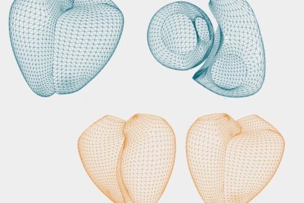
An artificial-intelligence software analyses heart scans in seconds and allows clinicians more time to spend with their patients.
News about the imaging project
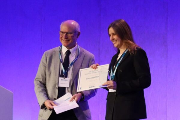
UK Biobank is thrilled to announce the winner of its inaugural Scientific Impact Awards, celebrating individuals and teams whose research is driving meaningful change in health through the innovative use of UK Biobank data.

The latest update to UK Biobank’s comprehensive dataset is now available to approved researchers around the world via UK Biobank’s Research Analysis Platform (UKB-RAP).

Most large studies typically scan just a single body part of a few thousand people, so this project is truly unique…not only are we working on a vastly bigger scale, but we record images of multiple parts of each person’s body, so you can study the whole person and see how it all relates.
Professor Sir Rory Collins, Chief Executive and Principal Investigator of UK Biobank