An Eye To The MRI
UK Biobank's imaging study aims to scan the brains, hearts, abdomens and bones of 100,000 participants and to repeat image 60,000. This imaging data will inform scientists on the subtle changes in our organs which can indicate ageing and age-related diseases, leading to earlier diagnosis, treatment and prevention. In our An Eye To The MRI series, we will showcase stories of researchers from the global scientific community who are using UK Biobank imaging data in their studies.
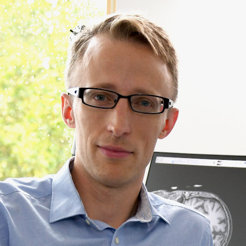
Dr Wenjia Bai: Imperial College London
Dr Wenjia Bai is a Senior Lecturer and an Associate Professor in the Department of Computing and Department of Brain Sciences, Imperial College London. His research focus has included analysing UK Biobank imaging data and developing automated analysis algorithms to understand aspects such as the shape, structure and function of our heart.
Wenjia is particularly interested in how artificial intelligence and deep learning techniques can be used to develop his automated image analyses to explore population-scale imaging data. He is excited to be able to use these enhanced analyses to ask questions over the next few years such as ‘are there any patterns in these images which are associated with the early onset of some diseases?’
‘The Biobank dataset is so unique…we have imaging scans which can give us very detailed descriptions of different organs in the body. This could create a huge opportunity for us to understand the risk factors for diseases.’
Professor Louise Thomas: University of Westminster
Louise Thomas is Professor of Metabolic Imaging at the University of Westminster and a Fellow of the Royal College of Physicians. Louise’s research is focused on measuring body composition using MRI. Her team has developed methods to quantify fat storage throughout the body and organs like the liver and to understand how this relates to disease risk.
Sponsored by numerous companies such as Calico and AMRA, she has used UK Biobank imaging data to develop faster methods for measuring organ volumes and fat distribution.
Louise is excited about the longitudinal aspect of UK Biobank; through our repeat imaging data, we can understand how bodies change as they age and what this means for our health. We caught up with Louise to find out more.
‘It allows us to look at very large populations and look at much smaller effects…UK Biobank has really revolutionised this area in collecting so much data.'
Dr James Cole: University College London
Meet Dr James Cole, an Associate Professor in Neuroimage Analysis, affiliated with the Dementia Research Centre and the Centre for Medical Image Computing, University College London.
James specialises in using magnetic resonance imaging (MRI) to observe how the brain changes as we age.
This research is crucial because with ageing comes an increased risk of age-associated diseases, many of which affect the brain. James has studied several of these brain diseases, from common diseases such as Alzheimer’s, to rarer conditions like Down’s Syndrome.
Ageing doesn’t affect everyone in the same way. James’s research illuminates why some people develop age-related diseases and others do not. This is important for the advancement of therapeutic treatments.
When James first started his PhD at King’s College London, observing the data of 30 or 40 people in a brain MRI study was considered ‘good’. The sheer size of UK Biobank’s imaging study today – tens of thousands of people – is “hugely important” in James’s eyes. With larger sample sets comes the opportunity to pinpoint patterns in results which may otherwise be hidden, and to usefully subdivide the data into smaller datasets to focus on a specific characteristic.
In addition, James highlights that “the way UK Biobank brings together different sources of information about the brain really is completely unique.” Indeed, the brain MRI data in UK Biobank is obtained through five or six types of brain scan. Capturing this depth of data in such a large number of people is extremely rare.
According to James, the most exciting aspect of UK Biobank’s imaging study is its longitudinal value.
Currently, participants are being invited back for their repeat scans, 2-7 years after their first. By comparing brain MRI data from the two scans, James can assess whether the way an individual’s brain looks in their first scan can be used to predict how the brain looks in their repeat scan. This will be highly valuable for accurately mapping how the brain changes in those with age-related diseases like dementia and ultimately, to the development of personalised medicine.

“Participants should be really proud of themselves for taking part in UK Biobank. Its imaging study is really quite unique in the world and will go on to add loads of great value for our understanding of ageing and health.”
~Dr James Cole
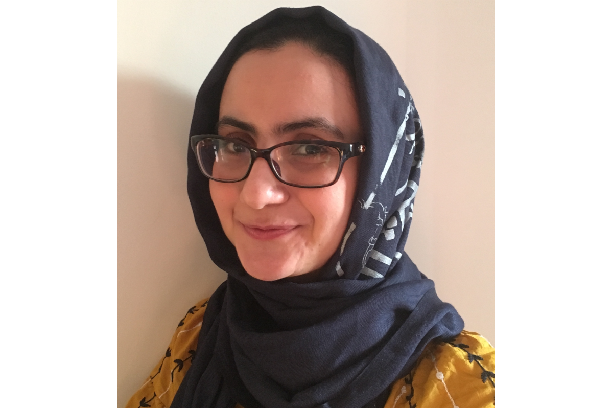
Dr Zahra Raisi-Estabragh: Queen Mary University of London

“The variety and quality of the data in UK Biobank allows us to address a very diverse range of important research questions. Without the altruism of its participants in giving their own time and effort, this depth of data would not exist."
~ Dr Zahra Raisi-Estabragh
Meet Dr Zahra Raisi-Estabragh, a National Institute of Health Research (NIHR) Clinical Lecturer at Queen Mary University of London and cardiology specialist at St Bartholomew's Hospital.
Zahra researches the causes of disease in different types of organs. She uses multi-organ imaging to capture changes in organs and spot diseases before symptoms have developed. For instance, detecting the first stages of heart disease in a seemingly healthy heart. These early insights may show how diseases develop and help clinicians to identify at-risk patients so that they can be treated earlier.
UK Biobank's large, highly-detailed imaging study is, according to Zahra, 'an amazing research resource.' The study accelerates researchers’ understanding of how imaging data can advance health prediction and disease prevention.
Zahra said that 'The variety and quality of the data in UK Biobank allows us to address a very diverse range of important research questions. Without the altruism of its participants in giving their own time and effort, this depth of data would not exist. UK Biobank is changing the way we do research into disease and our understanding of human health in a fundamental way; its value is huge and only going to become more significant as time goes on.'
It may sound like a small detail, but UK Biobank's 'standardised' format of MRI images allows researchers to accurately compare features between participants in different locations and across time. This is vital for our future plans to re-image people in a few years time and observe how their bodies have changed. We will be able to understand how quickly people are aging, and how rapidly changes, which may point towards disease, are occurring in different organs.
Last updated
